
Tissue method enables high quality imaging of heart cells

A new method for preparing ultra-thin slices of heart tissue in the lab could help scientists to study how cells behave inside a beating heart.
The heart is made up of millions of individual muscle cells called cardiomyocytes that are kept in place by a criss-crossing network of collagen fibres. These cells work together, contracting in sync to create a strong and regular heartbeat.
In patients with heart disease, however, these cells can start to die, beat out of sync, or the structure of the tissue can become damaged. In order to help patients with heart disease, researchers need to understand how and why these processes can go wrong and how they affect the organ.
Traditional methods for studying conditions affecting the heart have either used living animal models (such as rats and mice), which can be too complex to observe the heart in detail, or heart cells kept alive in the lab, which may provide an oversimplified view of what’s going on in the whole organ. A reliable intermediate model – which could be easily studied in the lab but without losing the complexity of the heart – has so far been out of reach.
Prepping the heart
Now, a team led by Professor Cesare Terracciano and Dr Filippo Perbellini, researchers at Imperial’s National Heart and Lung Institute, has perfected a method to reliably produce a much needed alternative, which can be used to prepare samples of heart tissue from small animals such as rodents, as well as larger animals and humans.
“The key difference with this approach is that we take the structure of the heart into account,” explains Dr Perbellini. “The heart is made up of sheets of muscle fibres, like a deck of cards. When we prepare the tissue we must do so in a way that aligns these sheets so that they remain intact and are not cut through.”

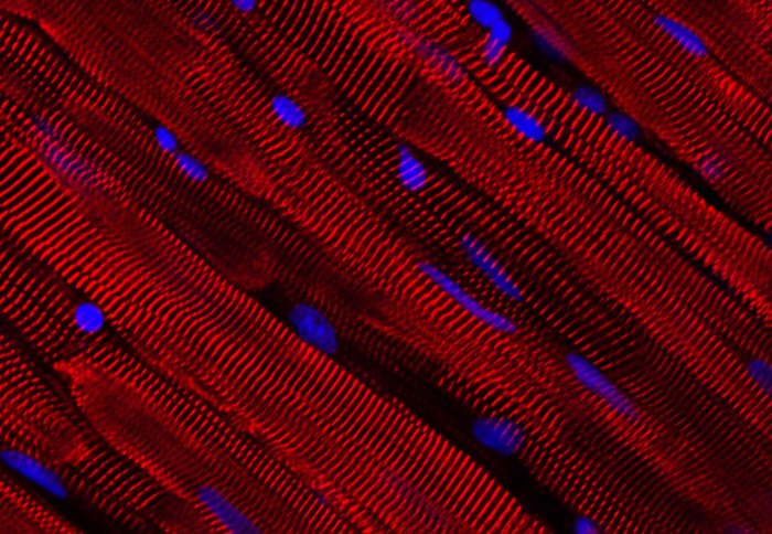
Tissue samples prepared using the method reveal calcium channels 'firing' in the heart muscle (white) fibres as the cells beat in unison

PhD researcher Samuel Watson, who outlined the procedure in a research paper this month, added: “Our method takes all of these factors into consideration and aligns the fibres so that the heart can be sliced without causing much damage.”
Researchers at Imperial College London were among the first to develop a technique which involves sheering off thin slices of tissue from the heart to study the organ.
According to Terracciano’s team, their refined method – which keeps more of the cells alive and the structure intact – could help to accelerate the development of new drugs and treatments for conditions such as coronary heart disease, inherited heart conditions, or heart failure, by providing a more accurate experimental model of the organ.
Better tool for testing drugs
For example, a tissue sample from the heart of a patient with heart failure could be removed soon after transplantation and used to test experimental drug treatments, showing which have the most beneficial effect on patients with the condition.
To obtain the samples, first the heart is cooled with an ice-cold solution before being removed from the donor’s chest and the tissue aligned before a precision cutting tool called a vibratome cuts the tissue, producing thin slices of living heart tissue that are less than half a millimetre thick. These sheets, between 100–400 µm in thickness – slightly thicker than a human hair – are cut from the walls of the heart chambers and provide an invaluable model for lab studies.
According to the researchers, the technique results in thin samples of tissue in which almost all of the cells (97%) are alive. These living sheets of tissue continue to contract, or ‘beat’, as they would in a living heart and far stronger than in traditionally prepared samples, as well as maintaining the intricate structure of the organ.
As the sheets are so thin, all of the cells are able to receive the oxygen and nutrients they need from a solution, so the tissue can be kept alive for longer – providing a tool to study heart disease. The researchers add that as multiple slices can be obtained from a single heart the approach could also help to reduce the numbers of animals used in heart research.
“Our laboratory was one of the first to use myocardial slices,” adds Professor Terracciano. “Through careful optimisation of the technique we have been able to improve it significantly, both in terms of increasing the proportion of living cells in the samples, as well as in terms of maintaining the structure and function of the tissue.”
The procedure is outlined in Nature Protocols, which features a high resolution image of a myocardial slice on the front cover.
-
‘Preparation of viable adult ventricular myocardial slices from large and small mammals’ by Samuel Watson et al. was published in Nature Protocols.
Article text (excluding photos or graphics) available under an Attribution-NonCommercial-ShareAlike Creative Commons license.
Photos and graphics subject to third party copyright used with permission or © Imperial College London.
Reporter

Ryan O'Hare
Communications Division
Contact details
Tel: +44 (0)20 7594 2410
Email: r.ohare@imperial.ac.uk
Show all stories by this author


Leave a comment
Your comment may be published, displaying your name as you provide it, unless you request otherwise. Your contact details will never be published.